New PET test may detect CTE in brains of living NFL players
Evidence of chronic traumatic encephalopathy, known as CTE, was found in 110 of 111 deceased NFL players’ brains that were donated to scientific research, according to medical journal by JAMA.

Ninety-nine percent.
The degenerative brain disease is associated with repeated head trauma, so football players would, logically, appear to be the most vulnerable to it. CTE can cause behaviors such as aggression, lack of impulse control, depression, suicidal thoughts and paranoia, and symptoms can begin in a patient’s late 20s or early 30s. The neurodegenerative disease has no treatment.
A study published in early 2019, done by a transnational team that included researchers from Arizona State University, Banner Alzheimer’s Institute, the Mayo Clinic College of Medicine and Boston University, revealed a step in the direction of identifying or developing examinations that could recognize CTE before death.
The study indicated an experimental positron emission tomography (PET) scan can detect an indication of CTE. The disease’s pathology is marked by a buildup of abnormal tau protein in the brain of living NFL players who have experienced cognitive, mood and behavioral symptoms. The researchers concluded the more years of tackle football played, the higher the tau protein level in the brain.
In 2017, Dr. Ann McKee, director of Boston University’s CTE center, said: "There's no question that there's a problem in football. That people who play football are at risk for this disease. And we urgently need to find answers for not just football players, but veterans and other individuals exposed to head trauma."
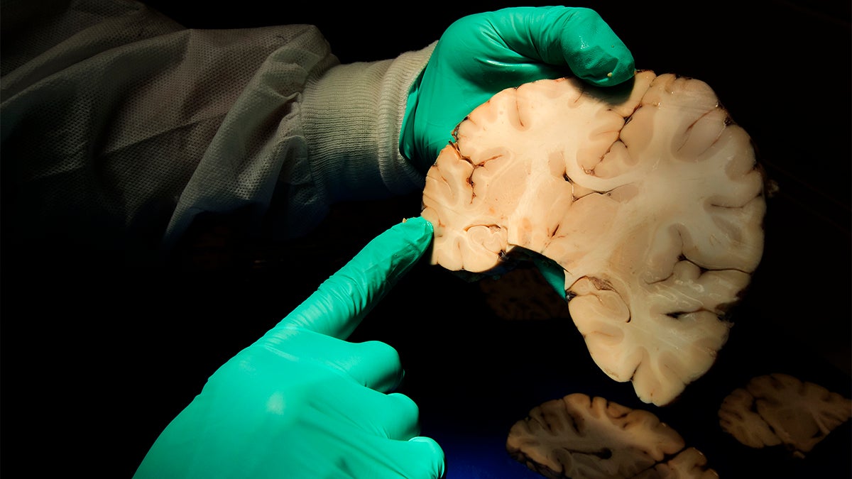
The problem with CTE is it cannot be detected without performing an autopsy, which leaves questions such as who has it, when will they get it or how will it affect their life. To confirm CTE, scientists and researchers must rely on players and families who agree to donate their brains to research after death.
“The results of this study provide initial support for the flortaucipir PET scan to detect abnormal tau from CTE during life. However, we’re not there yet,” Robert Stern, corresponding author, said. “These results do not mean that we can now diagnose CTE during life or that this experimental test is ready for use in the clinic.”
In CTE, a protein called tau forms clumps that slowly spread throughout the brain, killing brain cells as the protein spreads. The patient typically shows a lack of the amyloid plaques that are associated with Alzheimer’s disease.
A researcher with the ASU-Banner Neurological Disease Research Center at the Biodesign Institute, Diego Mastroeni, had a personal experience with this disease. A family member shared their concerns about memory issues, and how his NFL career may have affected his brain. Mastroeni was recruited to speak at Arizona’s NFL Alumni Association meeting.
“This was 2015,” Mastroeni said. “The NFL settlement was in full form, but players needed to be diagnosed with dementia or mild cognitive impairment in order to qualify. By this time, I was having personal communication with well over 50 retired NFL players. They were calling and emailing me daily, pleading for help, asking what to do.”
Diagnosing a living patient was very expensive and not reliable. A single brain scan cost around $100,000 at that time. Mastroeni talked about how vulnerable this population of males is, and how he knew he needed to find a way to help the players with no cost to them.
Though attempts to diagnose the disease during a person’s life had been unsuccessful, Mastroeni knew advances in brain imaging techniques, such as PET scans, were revolutionizing the field. These techniques already have provided a means to reliably detect and track brain changes in living people before the onset of memory loss in brain conditions, such as Alzheimer’s disease.
Mastroeni decided he needed help, so he reached out to neuroscientists Marwan Sabbagh and Eric Reiman and asked if they would be willing to talk with Mastroeni’s relative and others who had expressed their concern. The three connected and reached out to more people to add to their team.
Stern, who leads a CTE Center at BUSM, has been working to develop methods to detect and diagnose CTE in a living player. Stern continues to gain a better understanding of risk factors for the disease and works to understand why some players get it and some do not.
“A couple weeks later, Stern flew out from Boston, and we hashed out a plan to image all the guys we could,” Mastroeni said. “Some at BUSM, some at Banner and some at Mayo, at no cost to them.”
Mastroeni sent a letter to Arizona-based NFL players in 2014, offering them the chance to participate in a study. Although not all fit the criteria to participate, around 60 players responded, eager to be part of the study. The state of Arizona funded the study while researchers in Boston began their own recruiting and funding.
In the study, the researchers assessed amyloid plaque deposition in the brains of 26 living former NFL players with cognitive, mood and behavioral symptoms between the ages of 40 and 69. Researchers used experimental flortaucipir PET scans, which are used to measure tau levels in the brain, to do so. The experimenters used a control group of 31 men of the same ages who had no symptoms or history of traumatic brain injury.
The results showed tau PET levels were significantly higher in the former NFL player group than in the control group, and tau was seen in the specific areas of the brain that have been shown to be affected in post-mortem cases of neuro pathologically diagnosed CTE.
“Our findings suggest that mild cognitive, emotional and behavioral symptoms observed in athletes with a history of repetitive impacts are not attributable to AD, and they provide a foundation for additional research studies to advance the scientific understanding, diagnosis, treatment and prevention of CTE in living persons,” Reiman, a co-author, said. “More research is needed to draw firm conclusions, and contact sports athletes, their families, and other stakeholders are waiting.”
According to the report, the authors are partnering with the NIH and additional researchers to conduct a longitudinal study called the DIAGNOSE CTE Research Project in former NFL players, former college football players and persons without a history of contact sports play to help answers these questions. The results of this study are expected to be released in early 2020.
"The medical and scientific communities will benefit from this publication and the NFL will continue to work with a wide range of experts to improve the health of current and former NFL athletes," the NFL told CNN in a statement. "There are still many unanswered questions relating to the cause, incidence and prevalence of long-term effects of head trauma such as CTE."
Logan Huff is a senior journalism major at Arizona State University
Related Articles
The race to create the safest football helmet
Football takes a back seat to cycling when it comes to head injuries
Conflicting research on CTE shows need for more study
New research, technology aimed at minimizing concussions
Playing impact sports in high school can cause 'significant' changes in brain